Streamline detection with consistent, easy-to-read Northern and Southern blot transfers
Northern and Southern blotting are standard molecular biology techniques for identification and quantification of RNA and DNA, respectively. Effective isolation and detection of RNA and DNA in molecular biology research is critical to gene discovery, sequencing, and mapping used in diagnostics and industry applications.
Crucial to the blotting process is the clarity of the visual results
Northern and Southern blot analysis methods are based on the immobilization of nucleic acid. Nucleic acid fragments are separated by size onto a membrane to allow use of macromolecular probes for identification, detection, and visualization.
For unambiguous outcomes, to avoid re-work and delays, the transfer membrane used needs to have:
- The right level of sensitivity
- Low background noise
- Surface consistency
Northern blot transfers are used for the detection of specific RNA sequences among a mixture of diverse RNA. This technique can be used to characterize the expression of RNA from tissue samples or cell cultures. It involves a complex isolation and hybridization procedure which results in labeled probes bound to the RNA sequence of interest for detection and visualization.
Southern blotting is a molecular biology technique used for the detection of a specific DNA sequence in large, complex samples of DNA. The Southern blot method may also be used to determine the molecular weight of restriction fragments and to measure relative amounts of DNA in different samples. DNA samples can be obtained from tissue or isolated cell cultures.
Transfer membrane sensitivity can impact ease of detection
One of the challenges faced while identifying RNA or DNA fragments in samples through Northern and Southern blot analysis, is the relative abundance of RNA and DNA in the sample. When RNA and DNA molecules are less abundant, they can be difficult to isolate and visualize.
The membrane and detection protocol must provide the sensitivity to detect low numbers of molecules of interest without picking up background interference. A clear picture with low background is required to correctly quantify RNA expression patterns and overall DNA presence.
Cytiva supplies transfer membranes that demonstrate high sensitivity, low background, and lot-to-lot consistency for all radioactive and nonradioactive detection methods.
Northern blotting protocol
- The first step in Northern blotting requires isolation of RNA from biological samples.
- Once the RNA has been isolated, the RNA samples are separated by size via gel electrophoresis.
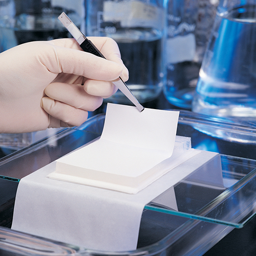
- The gel is blotted onto a nylon membrane, transferring and immobilizing the RNA onto the membrane.
- The membrane is incubated with short pieces of DNA (usually salmon sperm DNA) to block nonspecific interaction of the membrane material and detection probes.
- To identify the RNA, the membrane is placed in a hybridization buffer with a radioactively or chemically labeled probe specifically designed for the sequence of interest. This results in the hybridization of the probe to the RNA on the membrane blot that corresponds to the sequence of interest.
- Once hybridization is complete, the membrane is washed to remove any unbound probe.
- Finally, the labeled probe is detected via autoradiography or a chemiluminescent reaction, both of which result in the formation of a dark band on an X-ray film that can be used to compare expression patterns of the sequence of interest in the different samples.
Southern blotting protocol
- The first step in Southern blotting involves complete digestion of the DNA to be analyzed with a restriction enzyme.
- The complex mixture of DNA fragments is separated based on size by gel electrophoresis.
- The restriction fragments in the gel are denatured with alkali, making the DNA fragments accessible to the probe.
- The denatured DNA fragments are transferred onto a nylon or nitrocellulose membrane via blotting.
- The membrane is incubated with short pieces of DNA (usually salmon sperm DNA) to block nonspecific interaction of the membrane material and detection probes.
- Once the blocking is complete, the membrane is incubated under specific hybridization conditions with a specific radiolabeled DNA probe.
- Excess probe is washed from the probe bound to the membrane, and autoradiography is used to reveal the DNA fragment to which the probe hybridized.
Choosing the right membrane for your application
The Biodyne™ Nylon Transfer Membrane range (Pall™ Life Sciences products) offers a selection of chemistries with versatile adsorption properties for different applications. Our Biodyne A, Biodyne B, and Biodyne Plus membranes offer high sensitivity, low background, and lot-to-lot consistency for all radioactive and non-radioactive detection methods. These membranes are intrinsically hydrophilic resulting in easy wetting across the membrane. Their stability and durability ensure that they will not crack, shrink, or tear when subjected to multiple cycles of hybridization, stripping, and reprobing.
Biodyne A membrane: Amphoteric meaning the membrane zeta potential can be modulated by changes in pH. Well-suited for single probe or multiple rehybridizations, and applications where background is troublesome.
Biodyne B Membrane: Positively charged membrane. Our highest sensitivity nylon membrane for nucleic acid applications.
Biodyne Plus Membrane: Positively charged with an extremely high isoelectric point, which maintains its charge with changing pH. Suitable for chemifluorescent detection. With certain nonradioactive detection systems, it’s more sensitive than Biodyne A membrane while exhibiting lower background than Biodyne B membrane (refer to selection guide table in this article).
Biodyne C Membrane: Part of the Biodyne range, this membrane is applicable for specialist applications because it can be derivatized by coupling reactions through the carboxyl groups on the pore surfaces.
Table 1. Northern and Southern transfer membrane selection guide
| Product | Biodyne™ A Membrane | Biodyne B/Plus Membrane | Biodyne C Membrane |
| Description | Amphoteric nylon 6,6 | Positively-charged nylon 6,6 | Negatively-charged nylon 6,6 |
| Works best for: | Colony/plaque lifts, DNA and RNA transfers | DNA and RNA transfers, multiple reprobings | Reverse dot blots |
| Also suited for: | Gene probe assays, DNA fingerprinting, nucleic acid dot/slot blots, replica plating, ELISAs | DNA fingerprinting, Nucleic acid dot/slot blots, Colony/plaque lifts (Biodyne B membrane), Replica plating (Biodyne B membrane) | Protein immobilization, affinity purification, ELISAs |
| Advantages: |
• High sensitivity • Low background • Net change can be controlled by changing pH • Ability to strip and reprobe |
• Positive charge over broad pH range • Highest sensitivity for nucleic acid applications (Biodyne B membrane) • Ability to strip and reprobe |
• Negative charge over broad pH range • Surface carboxyl groups can be derivatized • Ability to strip and reprobe |
| Binding interaction: | Hydrophobic and electrostatic | Hydrophobic and electrostatic | Hydrophobic and electrostatic |
| Method of immobilization: | UV crosslink baking | Can be baked or UV crosslinked, although not required | Derivitization |
| Detection methods: | Radiolabeled probes, Enzyme-antibody conjugates
• Chemiluminescent • Chromogenic |
Radiolabeled probes, Enzyme-antibody conjugates
• Chemiluminescent • Chromogenic • Chemifluorescent (Biodyne Plus Membrane) |
Radiolabeled probes, Enzyme-antibody conjugates
• Chromogenic |
Explore our Northern and Southern blotting filtration products.

