Western blot protein analysis with clear results
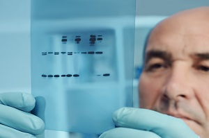
The Western blot (or immunoblot) technique has been a fundamental in protein analysis since the 1970s, when it was first discovered that biomolecules could be spotted directly onto membranes (spot ELISA or DNA dot blots), or transferred from gels (Southern blots, Northern blots, Western blots). Despite the now routine application of Western blotting, successful results can depend on several factors including optimal membrane selection for the desired detection method and protocol.
Western blotting is an analytical technique in molecular biology often used to investigate and characterize a protein’s post-translational modifications, for protein identification, and in protein production validation. Simple, yet effective, the Western blot has applications in many settings including basic science research, biopharmaceutical production, forensics, and diagnostics.
Achieve reliable and reproducible Western blot results
The key to reliable and reproducible Western blot results is a clean, low-background image. Problems can arise from:
- Membranes with low tensile strength
- High background signal
- High membrane burn-through
- Limited compatibility with detection methods
High-quality membranes are imperative to assess the relative abundance of proteins for analytical purposes. Our blotting membranes offer high tensile properties with low background levels. Our transfer membrane range supports a wide selection of detection methods without compromising quality.
The first step in a Western blot is the separation of proteins via gel electrophoresis from a sample containing a mixture of proteins. Our Nanosep™ centrifugal devices with low protein-binding Omega™ membranes (Pall™ Life Sciences products) are ideal to concentrate samples for gel electrophoresis.
Once separated on the gel, the proteins are transferred to a polyvinylidene fluoride (PVDF) membrane or a nitrocellulose membrane. This step immobilizes the proteins for further analysis.
The membrane is then incubated with labeled antibodies specific to the protein of interest.
After incubation, the membrane is washed to remove any unbound antibodies and further analyzed by various immunodetection techniques.
What is your Western blot detection method of choice?
The most commonly used detection methods involve radiolabels, fluorophores, and chromogenic and chemiluminescent enzymatic reactions. Each approach has its own benefits and considerations:
- Radiolabeled probes support protein detection directly via X-ray film; enhanced safety requirements are needed due to radioactivity.
- Chromogenic reactions do not need specialized detection equipment. Color-formation occurs on the membrane and can be visualized with the naked eye.
- Chemiluminescent reactions utilize light-sensitive equipment or materials to process and further analyze Western blot results.
- Fluorescent probes used as a detection method allow for multiplexing and concurrent detection of multiple proteins with different molecular weights.
Both qualitative and quantitative results rely on the difference between the background and the signal from the protein of interest.
Transfer membranes optimized for low noise and clear results
We offer a variety of transfer membranes that have been optimized for Western blotting applications (Table 1). The sensitive nature of our membranes yields high-resolution results with low background and burn-through.
- FluoroTrans™ PVDF transfer membrane has been optimized for fluorescent detection to ensure a very low background that will not interfere with protein detection and analysis when exposed to fluorescence. Autofluorescence from standard Western transfer membranes can obscure specific signals, especially at lower wavelengths, when using a fluorescent detection method.
- FluoroTrans™ W PVDF transfer membrane is optimal for use with traditional staining and chemiluminescent detection methods, as it has high sensitivity, low burn-through, and low background.
- BioTrace™ NT transfer membrane is a pure nitrocellulose unsupported membrane. It’s compatible with a variety of detection systems and has a high binding capacity for nucleic acids and proteins, thanks to the homogenous nature of the membrane. Other nitrocellulose membranes contain high levels of cellulose acetate, which reduces the protein binding capacity of the membrane. Physical characteristics of BioTrace™ NT membrane such as high tensile strength and hydrophilicity provide excellent handling and resistance to rips, tears, and cracking during transfers. Since the membrane is detergent free and unsupported, it yields sharper images with little to no distortion and has lower protein burn-through than competitive nitrocellulose media in electrophoretic transfers. Highly consistent, both inter- and intra- lot, this membrane provides sensitive detection of biomolecules blot after blot.
Table 1. Western blot transfer membrane selection guide
| Material | Nitrocellulose | PVDF | |
| Product | BioTrace™ NT | FluoroTrans™ PVDF | FluoroTrans™ W PVDF |
| Application | Colony/plaque lifts Western transfers Protein dot/slot blots |
(Primary) N-terminal protein sequencing (Also) Western transfers protein dot/slot blots |
(Primary)Western transfers (Also) N-terminal protein sequencing |
| Detection method | Radiolabeled probes Direct stain Fluorescence Enzyme-antibody conjugates Chemiluminescent Chromogenic |
Fluorescence Radiolabeled probes Direct stain Enzyme-antibody conjugates Chemiluminescent Chromogenic |
Radiolabeled probes Direct stain Enzyme-antibody conjugates Chemiluminescent Chromogenic |
| Features | No support fabric No detergents added 100% pure nitrocellulose |
Chemical resistance Non-flammable High strength Strong protein binding Very low burn-through Sensitive detection Good chemical compatibility |
Chemical resistance Non-flammable High strength Strong protein binding Very low burn-through Sensitive detection Good chemical compatibility |
| Benefits | Easily wetted with aqueous solutions, for less reagent usage Excellent strength |
Recommended for N-terminal protein sequencing: coating-free Low-fluorescence background suitable for fluorophore detection Highest protein affinity/avidity for optimal transfers |
Ideal for most Western blotting applications – except fluorophore detection High sensitivity; low background |
These are just a selection of the membranes we offer for Western blotting. Check page 6 of this selection guide for additional options.